BIaaS
NEXT gEnEration medical imaging
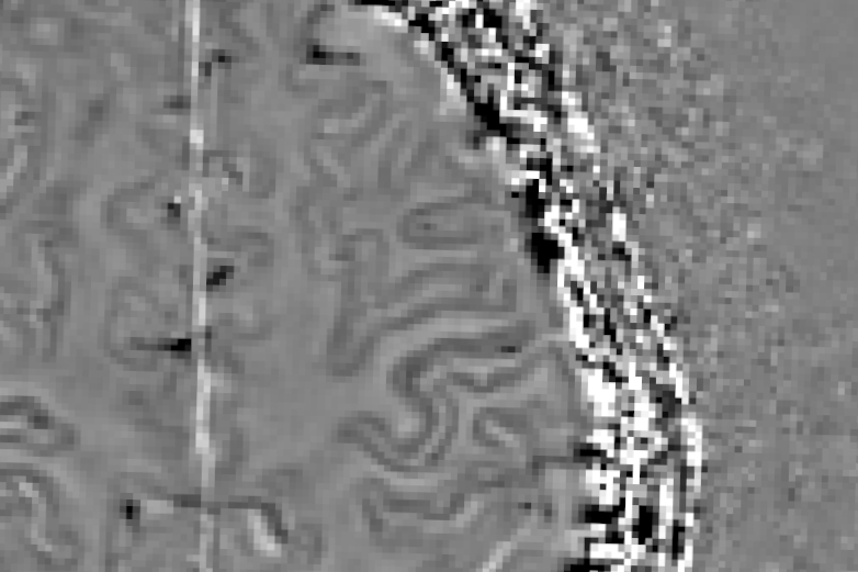
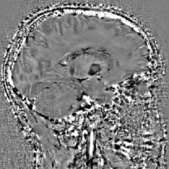
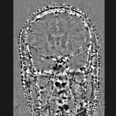
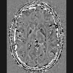
A robust and reliable mapping of iron distribution in the brain
investingating neurologic biomarkers
Iron: At the Heart of Brain Diseases
Brain iron plays a key role in many neurological disorders, but quantifying it remains complex and controversial.
Conventional imaging techniques provide poor detail about its precise distribution.
Our solutions enable the mapping of iron in vivo, providing reliable and reproducible measurements to explore its role in neurodegeneration.
MONITORING IRON LEVELS IN BRAIN DISEASES
ALZHEIMER
PARKINSON
VASCULAR DEMENCIA
MULTIPLE SCLEROSIS
NEURODEGENERATION WITH BRAIN IRON ACCUMULATION
HUNTINGTON DISEASE
TRAUMATIC BRAIN INJURY
ALZHEIMER
Alzheimer’s disease is a progressive neurodegenerative disorder that affects various regions of the brain.
The pathophysiological process involves the accumulation of Aβ peptides and tau proteins, along with changes in intracranial iron levels.
In addition to the protein biomarkers Aβ, Tau, and phosfoTau, PET-scan medical imaging allows for the visualization of Aβ peptide and Tau protein aggregates. Iron accumulations can be visualized and quantified without the injection of contrast agents using BIaaS (Brain-Iron-as-a-Service) MRI technology.
This quantification of iron via BIaaS in deep intracerebral regions is potentially linked to early prodromal stages of the disease. Our BIaaS solution could enable earlier detection and improve longitudinal patient monitoring. This technology is also of great interest for therapeutic monitoring and the monitoring of side effects from innovative therapies.
PARKINSON
Parkinson’s disease is a neurodegenerative disorder caused by the formation of aggregates of the α-synuclein protein within neurons.
In association with this aggregation, an accumulation of intracranial iron has also been demonstrated.
The diagnosis of this condition is generally based on symptoms such as resting tremors, limb rigidity, akinesia and gait disturbances.
Brain imaging, usually by MRI, is often limited to ruling out other causes. A brain scintigraphy scan may reveal the loss of dopaminergic neurons.
The quantification of intracerebral iron accumulation by MRI in the substantia nigra is well documented.
VASCULAR DEMENCIA
Vascular dementia is a neurodegenerative disease with multiple causes.
Damage to the brain’s nerve tissue may result from ischaemic lesions involving small blood vessels, multiple strokes, cognitive decline following a stroke, or a combination of these factors and another neurodegenerative disease such as Alzheimer’s.
As with many forms of dementia, diagnosis is based on clinical assessment of the patient, a gradual decline in cognitive function, and medical imaging.
BIaaS (Brain-Iron-as-a-Service), through its ability to detect microbleeds, can provide objective information on the aetiology of the disease.
MULTIPLE SCLEROSIS
Multiple sclerosis is an autoimmune disease characterised by lesions in the white matter, associated with chronic inflammation leading to demyelination.
An accumulation of iron has been observed in active, inflammatory lesions.
Diagnosis is based on the use of MRI to visualise and characterise the lesions. Nevertheless, the correlation between the number of lesions, their types and the severity of symptoms remains difficult to establish.
BIaaS (Brain-Iron-as-a-Service) enables the visualisation of iron accumulation within lesions and the categorisation of these lesions in terms of activity and progression. This solution could also contribute to the characterisation of the central vein sign (CVS).
The manifestation of iron, through the central vein sign and the paramagnetic halo, is now part of the international McDonald criteria for diagnosing and monitoring MS (revised version in September 2025).
NEURODEGENERATION WITH BRAIN IRON ACCUMULATION
Neurodegenerative diseases caused by intracerebral iron accumulation (NBIA) constitute a group of genetic disorders characterised by the progressive accumulation of iron in specific regions of the brain. Neuroferritinopathies and aceruloplasminemias are the two main categories of mutations, but there are numerous metabolic pathways involved in this family of diseases: lipid metabolism, mitochondrial function, lysosomal activity and autophagy.
Diagnosis is complex, the range of symptoms is wide and the pathophysiological processes are diverse.
Intracerebral iron accumulation is the common feature of this group of disorders. Quantifying this accumulation via MRI could be used as a method for diagnosis and monitoring of new therapies.
HUNTINGTON DISEASE
Huntington’s disease is a rare, hereditary neurodegenerative disorder. There is an adult-onset form and an even rarer juvenile form. It is characterised by motor and cognitive impairments.
The diagnosis of Huntington’s disease is based on family history and characteristic symptoms. PET and MRI scans enable a differential diagnosis to be made and, in particular, allow assessment of atrophy of the caudate and putamen.
A significant increase in iron has been observed in patients in the caudate, putamen and globus pallidus. BIaaS could facilitate the monitoring of the disease and the development of new therapies.
TRAUMATIC BRAIN INJURY
Traumatic brain injuries encompass a range of neurological disorders that may result from direct impact, acceleration-deceleration forces, intracranial haemorrhages or diffuse axonal injury. These conditions can lead to cognitive decline, functional impairments and, in some cases, late-onset neurodegenerative changes.
Diagnosis is based on clinical assessment, changes in cognitive function and imaging, particularly CT or MRI scans. Symptoms may progress in stages depending on the development of secondary lesions, such as microhaemorrhages.
BIaaS (Brain-Iron-As-A-Service), thanks to its ability to detect veins, haemorrhages and microbleeds, could provide more accurate objective information on the nature and extent of post-traumatic lesions.
A 100% integrated and compatible solution
Compatible on 100% MRI systems
Access via a secure cloud-based SaaS solution
PACS - DICOM compatible
Medical device marking pending
Indications and Applications
Preclinic studies
Clinic studies
Medical device
Marking as a medical device pending
They trust us